- Home
- Diabetes Complications
- What Is the Best Treatment for Diabetic Retinopathy
Diabetic Retinopathy Treatment Options: Injections, Laser and Surgery
When I tell a patient that diabetes has affected the retina, one of the first questions I hear is, “Will I lose my sight?” This fear is understandable, but a diagnosis of diabetic retinopathy does not mean that blindness is inevitable. Modern treatment can often stabilize the retina, reduce swelling, control abnormal blood vessels, and protect useful vision—especially when the problem is found early and follow-up appointments are kept.
The best treatment is not the same for every eye. An ophthalmologist, often a retina specialist, chooses treatment according to the stage of retinopathy, whether the macula is swollen, how clearly you can see, whether abnormal vessels are growing, and whether bleeding or scar tissue is pulling on the retina. One eye may need treatment while the other only needs observation.
Treatment Starts With an Accurate Retinal Diagnosis
Diabetic retinopathy damages the tiny blood vessels of the retina, the light-sensitive tissue at the back of the eye. In earlier nonproliferative diabetic retinopathy (NPDR), vessels may weaken, develop small bulges, leak, or become blocked. In proliferative diabetic retinopathy (PDR), the oxygen-starved retina releases signals that encourage fragile new vessels to grow. These vessels can bleed into the vitreous gel and create scar tissue that pulls on the retina.
Diabetic macular edema (DME) is swelling in the macula, the central part of the retina used for reading, recognizing faces, and seeing fine detail. DME can occur at any stage of retinopathy. It is important to distinguish it from PDR because the treatment goal and method may be different.
A dilated retinal examination is the foundation of diagnosis. The specialist may also use optical coherence tomography (OCT) to measure retinal swelling, retinal photography to document changes, and fluorescein angiography or OCT angiography to assess leaking, blocked, or abnormal vessels. Visual-acuity testing helps determine whether swelling is affecting function. Treatment should be based on the complete eye examination—not on symptoms or an HbA1c result alone.
Early retinopathy does not always need an immediate procedure
When NPDR is mild or moderate and there is no vision-threatening macular edema, careful observation may be the safest plan. “Observation” does not mean ignoring the condition. It means returning at the interval recommended by the eye specialist, completing retinal imaging when needed, and improving modifiable risks. Some people need annual review, while more advanced changes may require visits every few months.
Which Treatment Is Used at Each Stage?
| Eye finding | Common management approach | Main purpose |
|---|---|---|
| Mild or moderate NPDR without vision-threatening DME | Monitoring, retinal imaging when indicated, and management of systemic risk factors | Detect progression before vision-threatening complications develop |
| Center-involving DME with reduced vision | Anti-VEGF injections are commonly first-line; steroid or laser treatment may be appropriate in selected eyes | Reduce macular swelling and protect or improve central vision |
| Center-involving DME with good vision | Careful observation, anti-VEGF treatment, or laser may be considered depending on anatomy and specialist judgment | Avoid unnecessary treatment while responding promptly if vision worsens |
| Proliferative diabetic retinopathy | Panretinal photocoagulation, anti-VEGF injections, or a planned combination | Control fragile new vessels and reduce the risk of severe bleeding or retinal detachment |
| Non-clearing vitreous hemorrhage, tractional detachment, or extensive scar tissue | Vitrectomy, sometimes with laser and other procedures during surgery | Clear blood, relieve traction, and repair or stabilize the retina |
This table is a guide, not a prescription. The exact plan depends on both eyes, vision, OCT findings, pregnancy, previous treatment, other eye diseases, ability to attend frequent appointments, access and cost, and the ophthalmologist’s assessment.
Anti-VEGF Eye Injections
Vascular endothelial growth factor, or VEGF, is a protein involved in leakage and the growth of fragile abnormal retinal vessels. Anti-VEGF medicines block this signal. They are not weak or merely temporary medicines; they have transformed the treatment of diabetic macular edema and proliferative diabetic retinopathy.
Common anti-VEGF medicines include aflibercept, ranibizumab, faricimab, and bevacizumab. Availability, licensing, insurance coverage, and use differ by country. Bevacizumab is frequently used off-label in ophthalmology. Your retina specialist considers visual acuity, OCT findings, previous response, treatment interval, cost, pregnancy status, and other health factors when selecting a medicine.
What happens during an injection?
The eye is numbed, the eyelids and surrounding area are cleaned carefully, and a small device may hold the eyelids open. Medicine is injected into the vitreous through the white part of the eye. The injection itself is usually brief. A sensation of pressure, mild irritation from the antiseptic, a small red area on the white of the eye, or temporary floaters may occur afterward.
Many people need frequent injections at first. The interval may later be extended if the macula becomes drier and the disease remains controlled. Others need ongoing treatment. Missing visits matters: VEGF activity can return, swelling may recur, and abnormal vessels can become active again. Anti-VEGF treatment controls disease; it does not remove diabetes or guarantee a permanent cure.
Benefits and risks
Anti-VEGF treatment can reduce retinal swelling, improve vision in some eyes, preserve vision in many others, and cause abnormal vessels to regress. Rare but serious eye risks include infection inside the eye, retinal tear or detachment, bleeding, inflammation, and a significant rise in eye pressure. Your specialist will also consider your wider medical history when discussing possible systemic risks.
Seek immediate advice for worsening eye pain, increasing redness, marked light sensitivity, pus or discharge, rapidly worsening floaters, or decreasing vision. These symptoms can indicate infection or another complication and should not wait for the next routine appointment.
Corticosteroid Injections and Implants
Corticosteroids reduce inflammation and retinal leakage. They may be delivered as an injection or a longer-acting implant. Steroids are not used simply to “stop bleeding,” and they are not automatically combined with surgery. They are mainly considered for DME in selected situations—for example, when anti-VEGF treatment has not produced an adequate response, when frequent anti-VEGF visits are difficult, or when an ophthalmologist believes the balance of benefits and risks favors a steroid.
The important trade-offs are cataract formation or progression and increased pressure inside the eye, which can lead to glaucoma. Eye pressure must therefore be monitored. The choice may differ between an eye that still has its natural lens and one that has already had cataract surgery. Steroids may reduce swelling effectively, but follow-up remains essential.
Laser Treatment: Two Different Purposes
The word “laser” can describe different procedures. Laser is not a single treatment that permanently seals every leaking blood vessel. The retina can continue to change, so further treatment or injections may still be needed.
Focal or grid laser for diabetic macular edema
Focal/grid laser places small burns around selected areas of leakage away from the center of the macula. It may be considered for non-center-involving DME, persistent edema, or particular leaking lesions. For center-involving DME that has reduced vision, anti-VEGF therapy has largely replaced laser as the usual initial treatment, although laser can still have a supporting role.
Panretinal photocoagulation for proliferative retinopathy
Panretinal photocoagulation (PRP), also called scatter laser, treats many areas of the peripheral retina. Its purpose is to reduce the drive for abnormal vessel growth and lower the risk of severe bleeding and vision loss. It has protected sight for decades and remains an important, durable treatment for PDR.
PRP may require more than one session. It can affect night vision, peripheral vision, and color vision, and it may worsen existing macular edema in some eyes. Anti-VEGF treatment can preserve peripheral vision better, but usually demands more frequent injections and dependable follow-up. For a person who may not be able to return reliably, the relative durability of PRP can be especially important. Some patients benefit from both approaches.
Vitrectomy Surgery
A vitrectomy removes vitreous gel and blood from inside the eye through very small openings. The surgeon can also remove scar tissue, relieve traction pulling on the retina, apply laser, repair retinal detachment, and place air, gas, or silicone oil in the eye when needed.
Vitrectomy may be recommended for:
- dense or persistent vitreous hemorrhage that blocks vision or prevents examination and treatment of the retina;
- tractional retinal detachment threatening or involving the macula;
- combined tractional and retinal-tear detachment;
- extensive scar tissue or continued severe PDR despite other treatment;
- selected cases of DME associated with traction on the macula.
Vitrectomy can clear the visual pathway and stabilize anatomy, but the final vision depends on how healthy the macula and optic nerve remain. Long-standing swelling, poor retinal blood flow, or previous detachment can limit recovery. Risks include cataract, infection, bleeding, pressure changes, recurrent detachment, and the need for more surgery.
If a gas bubble is placed in the eye
Follow the surgeon’s positioning instructions exactly. Do not fly or travel to high altitude until the surgeon confirms that the bubble is gone. Tell every doctor and anesthetist that you have an eye gas bubble because nitrous-oxide anesthesia can cause a dangerous pressure rise. Your own surgical team’s instructions take priority.
Does Cryotherapy Still Treat Diabetic Retinopathy?
Retinal cryotherapy applies freezing treatment through the outside wall of the eye. It was used more often before modern laser systems, anti-VEGF medicines, and small-incision vitrectomy became widely available. It is not a routine first-line treatment for diabetic macular edema and is not used simply to shrink all diabetes-damaged vessels.
A retinal surgeon may consider cryotherapy in uncommon, complex circumstances—for example, when the retina cannot be treated adequately with laser because the view is blocked or when a specific surgical problem requires it. Its role is specialized. A patient should not expect cryotherapy merely because retinopathy or retinal swelling has been diagnosed.
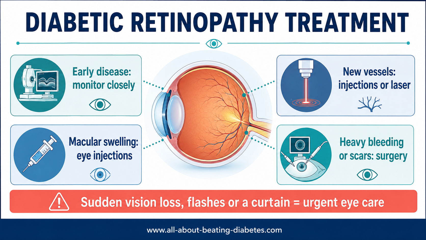

How Your Retina Specialist Chooses a Treatment
Two people with “diabetic retinopathy” may need very different care. I encourage patients to ask which exact finding is being treated. Is it center-involving macular edema? New abnormal vessels? Bleeding? Scar tissue? A threatened detachment? Understanding this makes the plan much clearer.
The decision may consider:
- NPDR or PDR stage and whether it is changing;
- presence, location, and thickness of macular edema on OCT;
- current vision and the vision in the other eye;
- previous response to injections, steroid, or laser;
- cataract, glaucoma, eye-pressure history, or previous eye surgery;
- pregnancy or plans for pregnancy;
- kidney disease, cardiovascular history, and other medical factors;
- ability to return for repeated treatment and monitoring;
- medicine availability, insurance coverage, and cost.
If repeated injections are recommended, ask what improvement the specialist hopes to see, how response will be measured, and what would lead to extending the interval, switching medicine, adding laser, or considering surgery. If PRP is recommended, ask about the expected benefit, the number of sessions, and possible effects on night or peripheral vision.
What to Expect From Follow-Up
Retinopathy can remain active even when vision feels unchanged. Keep every scheduled visit unless the clinic advises otherwise. Your eye team may repeat vision testing, pressure measurement, dilated examination, photography, or OCT. These results show whether swelling is improving, new vessels are inactive, bleeding is clearing, or traction is changing.
After laser, vision may be temporarily blurry and the eye may feel sensitive. Arrange transport if your clinic recommends it, and follow instructions about driving. After surgery, use drops as directed, avoid rubbing the eye, follow positioning and activity restrictions, and attend all postoperative checks. Do not stop prescribed treatment because the eye feels better.
Glasses do not treat retinopathy, although an updated prescription may help once glucose and retinal status are stable. Herbs, vitamins, eye exercises, and dietary supplements do not replace retinal injections, laser, surgery, or medical follow-up. If visual damage cannot be fully corrected, low-vision rehabilitation, magnification, lighting changes, phone accessibility tools, and mobility support can preserve independence.
Protecting the Retina Beyond Eye Procedures
Eye treatment and diabetes treatment work together. Managing blood glucose reduces the risk of retinopathy developing and progressing, but the goal should be individualized and safe. Blood pressure, cholesterol, smoking, kidney disease, anemia, pregnancy, and sleep apnea may also influence retinal risk. Review these factors with your diabetes team.
Do not make a sudden, large medication change by yourself to lower HbA1c rapidly. In some people with existing retinopathy, rapid improvement in severe hyperglycemia can temporarily worsen retinal disease. This does not mean that high glucose should remain untreated. It means glucose improvement and eye monitoring should be coordinated when retinopathy is advanced or HbA1c is very high.
If you have type 2 diabetes, a comprehensive eye examination is generally recommended at diagnosis because diabetes may have been present silently for years. In type 1 diabetes, screening usually begins within several years of onset. Pregnancy with pre-existing diabetes requires early and often closer eye monitoring. Once retinopathy is present, the ophthalmologist—not a fixed online timetable—should set the follow-up interval.
Doctor’s Note from Dr. Albana Greca
Do not wait for pain to tell you that retinopathy is getting worse. The retina often changes without pain and before you notice vision loss. The most protective steps are simple to state but important to follow: attend dilated eye examinations, know the exact retinal diagnosis, keep injection or laser appointments, and coordinate eye care with glucose, blood-pressure, cholesterol, and kidney care.
When Vision Symptoms Need Urgent Care
- sudden loss or marked worsening of vision;
- a sudden shower of new floaters or cobweb-like spots;
- flashes of light;
- a dark curtain, shadow, or missing area in the visual field;
- new distortion or a sudden central blind spot;
- severe eye pain, headache, nausea, or a red eye with reduced vision;
- worsening pain, redness, or vision after an injection or eye operation.
These symptoms may indicate vitreous hemorrhage, retinal tear or detachment, acute glaucoma, infection, vascular blockage, or another emergency. Do not assume every visual change is “just high sugar,” and do not drive yourself if vision is unsafe.
Frequently Asked Questions
Can diabetic retinopathy be cured permanently?
Current treatments can control leakage, swelling, abnormal vessels, bleeding, and traction, but they do not remove diabetes or guarantee that the condition will never return. Continued retinal follow-up and management of glucose, blood pressure, cholesterol, and other risks remain necessary.
Can vision improve after treatment?
Yes, especially when vision loss is caused by treatable macular swelling or blood blocking the visual pathway. However, improvement is not guaranteed. Long-standing macular damage, poor retinal circulation, scar tissue, retinal detachment, or optic-nerve damage can limit recovery. Treatment may still be valuable because preventing further loss is an important success.
Are eye injections painful?
The eye is numbed before treatment, so most people report pressure or brief discomfort rather than severe pain. Antiseptic can cause irritation afterward. Worsening pain, redness, light sensitivity, or reduced vision after an injection requires urgent contact with the eye clinic.
How many anti-VEGF injections will I need?
There is no fixed number for everyone. Injections are often frequent at the beginning and may later be spaced farther apart according to vision, OCT results, and disease activity. Some people can pause treatment under monitoring; others need continuing injections to protect vision.
Is laser better than eye injections?
Neither is universally better. Anti-VEGF is commonly first-line for center-involving DME with vision loss. For PDR, anti-VEGF and PRP are both effective options with different advantages. PRP can be more durable but may affect peripheral and night vision; injections can preserve more peripheral vision but require reliable repeated follow-up.
Does laser stop retinal leaking forever?
No. Laser can reduce leakage or suppress the stimulus for abnormal vessels, depending on the laser type, but diabetic retinal disease can remain active or recur. Some people need repeat laser, injections, surgery, or continued observation.
When is vitrectomy necessary?
Vitrectomy may be recommended for dense or non-clearing vitreous hemorrhage, tractional retinal detachment, extensive scar tissue, combined retinal detachment, or selected traction-related macular problems. A retina surgeon decides whether the likely benefit outweighs surgical risk.
Can better blood sugar replace eye treatment?
No. Safer glucose control can slow future damage, but it cannot reliably remove active macular edema, abnormal new vessels, major bleeding, or scar traction. If your ophthalmologist recommends injections, laser, or surgery, do not delay it while trying diet, supplements, or glucose improvement alone.
Related Questions
Related Resources
References
- National Eye Institute: Diabetic Retinopathy.
- National Eye Institute: Injections to Treat Eye Conditions.
- American Academy of Ophthalmology: Diabetic Retinopathy Preferred Practice Pattern.
- American Diabetes Association: Retinopathy, Neuropathy, and Foot Care—Standards of Care in Diabetes 2026.
- NICE: Diabetic Retinopathy—Management and Monitoring.
- National Eye Institute: Retinal Detachment and Emergency Symptoms.