- Home
- Diabetes Complications
- Diabetic Eye Problems Symptoms and Treatment
Diabetic Eye Problems: Symptoms, Screening, Treatment, and Emergency Signs
This article explains how diabetes can affect the eyes, the warning signs to watch for, and the importance of regular eye examinations for preventing vision loss.
Written by Dr. Albana Greca, MD, MMedSc, Family Physician
Medically reviewed by Dr. Ruden Cakoni, MD, Endocrinologist
Last reviewed: July 2026
Quick Answer
Diabetes can affect the retina, macula, lens, optic nerve, eye muscles, and the eye’s drainage system. The main diabetes-related eye conditions are diabetic retinopathy, diabetic macular edema, cataracts, and glaucoma.
Early diabetic retinopathy often causes no symptoms, so a comprehensive dilated eye examination is essential even when vision seems normal. Sudden vision loss, a dark curtain over the vision, a sudden shower of floaters or flashes, severe eye pain, or new double vision requires urgent medical assessment.
Diabetic eye disease is not one single condition. It is a group of problems that can affect different structures of the eye. Some changes are temporary, such as short-term blurry vision caused by rapidly changing glucose levels. Others can permanently threaten sight if they are not detected and treated early.
The original version of this page focused mainly on direct optic-nerve damage and inflammation. In practice, the most characteristic diabetes-related eye disease begins with damage to the small blood vessels of the retina. Diabetes also increases the risks of cataracts and glaucoma, and it can occasionally affect nerves that control eye movement.
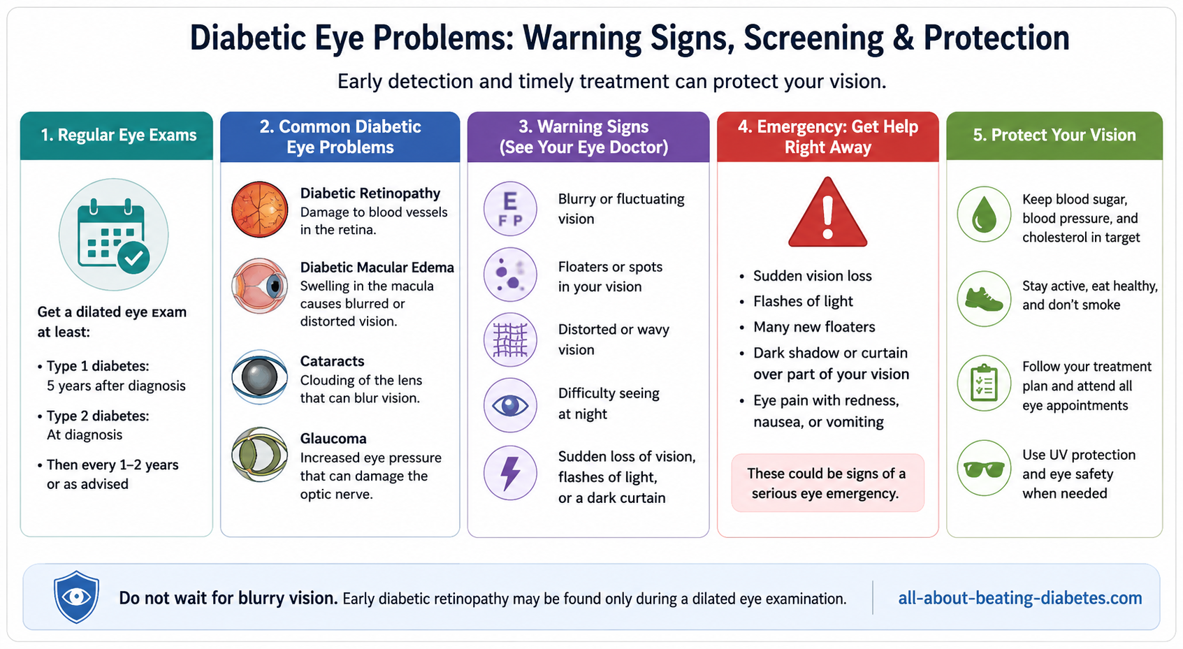

The Main Diabetic Eye Problems
1. Diabetic retinopathy
The retina is the light-sensitive layer at the back of the eye. It converts light into signals that travel through the optic nerve to the brain.
Over time, elevated glucose, high blood pressure, and other vascular risk factors can damage the retina’s small blood vessels. The disease usually progresses through two broad stages:
- Nonproliferative diabetic retinopathy: retinal blood vessels may weaken, bulge, close, or leak blood and fluid.
- Proliferative diabetic retinopathy: the oxygen-deprived retina stimulates fragile new blood vessels. These vessels can bleed, scar, and pull the retina away from the back of the eye.
Retinopathy can be mild for years without affecting vision. That does not mean it should be ignored. The stage, location, and rate of progression determine how often the eyes need monitoring and whether treatment is required.
2. Diabetic macular edema
The macula is the central part of the retina used for reading, driving, recognizing faces, and seeing fine detail. When damaged retinal vessels leak fluid into this area, the macula can swell. This is called diabetic macular edema, or DME.
DME can occur at any stage of diabetic retinopathy and may cause blurry, distorted, or wavy central vision. It is a major cause of vision loss in diabetes.
3. Cataracts
A cataract is clouding of the eye’s natural lens. People with diabetes are more likely to develop cataracts and may develop them at a younger age.
Possible symptoms include:
- Cloudy or blurred vision
- Glare or halos around lights
- Difficulty driving at night
- Faded colors
- Frequent changes in glasses prescription
- Double vision in one eye
Learn more from our guide to cataracts and type 2 diabetes.
4. Glaucoma
Glaucoma damages the optic nerve. Open-angle glaucoma often progresses slowly without early symptoms and gradually reduces peripheral vision. Diabetes increases the risk of glaucoma.
Advanced diabetic retinopathy can also cause abnormal vessels to block the eye’s drainage angle. This form, called neovascular glaucoma, may cause severe eye pain, redness, headache, nausea, halos, and sudden vision loss.
5. Temporary blurry vision from changing glucose
High or rapidly changing glucose can alter fluid levels in the lens and focusing tissues. Vision may become blurry for days or weeks while glucose is changing. This type of blur may improve when glucose stabilizes.
Do not assume that all blurry vision is temporary. Persistent, recurrent, one-sided, or sudden blurring needs an eye examination. Read our broader explanation of blood glucose symptoms and patterns.
6. Double vision and diabetes-related nerve palsy
Double vision means seeing two images of one object. Diabetes can occasionally affect one of the nerves that controls eye movement, causing sudden binocular double vision, eye misalignment, or a drooping eyelid.
Double vision can also result from stroke, aneurysm, thyroid eye disease, myasthenia gravis, cataract, or another neurological or eye condition. New double vision should be assessed promptly—especially with weakness, facial drooping, speech difficulty, severe headache, or unequal pupils.
Symptoms of Diabetic Eye Disease
There may be no early symptoms. When symptoms develop, they can include:
- Blurred or fluctuating vision
- Wavy or distorted central vision
- Difficulty reading or recognizing faces
- Dark areas or missing areas in the visual field
- Floaters, spots, cobwebs, or dark strings
- Flashes of light
- Poor night vision
- Reduced contrast or faded colors
- Loss of peripheral vision
- Double vision
- Eye pain, redness, headache, or halos
The old claim that blurry or double vision is the first symptom in 80% of patients was unsupported. Many people with clinically important retinopathy have no symptoms until the disease is advanced.
Are black spots always advanced retinopathy?
No. Floaters can occur because of normal age-related changes, posterior vitreous detachment, inflammation, bleeding, or retinal tears. In a person with diabetes, a sudden increase in dark spots or cobwebs may indicate vitreous bleeding or a retinal tear and needs urgent assessment.
Does eye pain mean inflammation?
Not necessarily. Diabetic retinopathy is often painless. Eye pain can result from acute glaucoma, corneal disease, infection, inflammation, migraine, sinus disease, or another condition. Severe pain with redness, headache, nausea, halos, or vision loss is an emergency.
When to Seek Urgent or Emergency Eye Care
Seek same-day urgent eye assessment for:
- A sudden shower of new floaters
- New flashes of light
- A dark curtain, veil, or shadow over part of the vision
- Sudden or rapidly worsening vision loss
- New double vision
- New eye pain or marked redness
- Blood appearing inside the visible part of the eye
Call emergency services immediately for:
- Vision loss with facial drooping, weakness, speech trouble, severe imbalance, or confusion
- Severe headache with new double vision, unequal pupils, or a drooping eyelid
- Severe eye pain with vomiting or sudden major vision loss
- Eye trauma or a chemical injury
When Should People With Diabetes Have Eye Exams?
| Situation | Recommended Initial Examination | Follow-Up |
|---|---|---|
| Type 1 diabetes | Comprehensive dilated eye examination within 5 years after diabetes begins | Usually annually; frequency depends on findings and risk |
| Type 2 diabetes | At diagnosis | Usually annually; some people with repeated normal exams and stable glucose may be advised to return every 1–2 years |
| Existing retinopathy | Prompt ophthalmology assessment | At least annually and often more frequently, depending on severity and progression |
| Pregnancy with preexisting type 1 or type 2 diabetes | Before pregnancy when possible or during the first trimester | Each trimester and after delivery as advised, based on retinopathy severity |
“Annual” is a general guide. Someone with proliferative retinopathy, macular edema, rapidly changing vision, pregnancy, kidney disease, or rapidly worsening glucose may need examinations every few weeks or months.
Can retinal photographs replace an eye examination?
High-quality retinal photography with remote expert interpretation or validated artificial-intelligence systems can improve access to screening. It is not a complete substitute when images are poor, symptoms are present, disease is detected, eye pressure must be measured, or another eye condition is suspected. Abnormal results require referral to an eye-care professional.
How Diabetic Eye Problems Are Diagnosed
A comprehensive diabetes eye assessment may include:
- Visual acuity testing
- Dilated examination of the retina and optic nerve
- Slit-lamp examination
- Measurement of eye pressure
- Optical coherence tomography, or OCT, to measure retinal and macular swelling
- Retinal photography
- Fluorescein angiography when detailed blood-vessel imaging is needed
- Visual-field testing for glaucoma or neurological disease
- Ultrasound when bleeding prevents a clear view of the retina
A routine glasses examination without dilation may not be sufficient to exclude diabetic retinopathy.
Treatment of Diabetic Eye Disease
Treatment depends on the exact condition, stage, vision, retinal anatomy, other eye disease, pregnancy, ability to attend follow-up, and overall health. Early retinopathy may need monitoring rather than immediate eye procedures.
Anti-VEGF eye injections
Vascular endothelial growth factor, or VEGF, promotes leakage and abnormal blood-vessel growth. Anti-VEGF medicines are injected into the eye after numbing and sterile preparation.
They are commonly used for center-involving diabetic macular edema that affects vision and can also be used to treat proliferative diabetic retinopathy. Several injections may be required initially, followed by individualized monitoring and treatment.
Examples include aflibercept, ranibizumab, bevacizumab, and faricimab. The exact choice depends on local approval, vision, OCT findings, treatment response, cost, and the ophthalmologist’s recommendation.
Laser treatment
Laser remains important but is no longer the only central treatment.
- Panretinal photocoagulation: treats proliferative retinopathy by reducing the stimulus for dangerous new blood vessels.
- Focal or grid laser: may be used for selected cases of macular edema, particularly when leakage is outside the center or edema persists.
Laser can reduce the risk of severe vision loss, but it does not guarantee that lost vision will return. Panretinal laser can sometimes reduce peripheral or night vision.
Intravitreal corticosteroids
Corticosteroid injections or implants may be considered for selected eyes with persistent diabetic macular edema, particularly when anti-VEGF treatment is unsuitable or insufficient.
They are not the routine treatment for every diabetic eye problem. Important risks include increased eye pressure, glaucoma, and cataract. The old page discussed general corticosteroid side effects such as weight gain and bone loss; those are more relevant to systemic steroids than to the carefully selected local eye treatments used by retina specialists.
Vitrectomy surgery
Vitrectomy may be needed for:
- Vitreous hemorrhage that does not clear
- Tractional retinal detachment threatening or affecting the macula
- Dense scar tissue
- Some severe cases of proliferative retinopathy
- Selected cases of persistent macular edema with traction
Surgery can preserve or improve vision, but it cannot always restore normal sight or prevent future disease. Ongoing retinal monitoring and systemic diabetes care remain necessary.
Cataract and glaucoma treatment
Cataract surgery replaces the cloudy lens with an artificial lens. The final visual result also depends on the health of the retina, macula, and optic nerve.
Glaucoma treatment may include pressure-lowering eye drops, laser, injections for neovascular disease, or surgery. Severe painful neovascular glaucoma requires urgent specialist care.
Our separate diabetic retinopathy treatment guide provides a more focused explanation of injections, laser, and retinal surgery.
How to Reduce the Risk of Vision Loss
Manage glucose safely
Keeping glucose and HbA1c near individualized targets can reduce the risk of retinopathy and slow progression. However, abrupt major improvement in glucose can temporarily worsen retinopathy in some people who already have eye disease.
Do not avoid needed glucose treatment. Instead, tell the prescribing clinician about known retinopathy and keep the planned eye follow-up when diabetes therapy is intensified. Our HbA1c guide explains how long-term glucose is assessed.
Control blood pressure and cholesterol
High blood pressure and abnormal cholesterol increase vascular risk. Take prescribed medicines and attend routine monitoring.
Do not smoke
Smoking contributes to vascular damage and increases overall cardiovascular and eye-health risk.
Keep kidney and pregnancy care coordinated
Diabetic kidney disease and retinopathy often occur together because both involve small-vessel injury. Kidney disease, pregnancy, and rapidly changing glucose can justify closer eye monitoring. See our diabetic kidney disease guide.
Attend every appointment
Anti-VEGF treatment and monitoring schedules matter. Missing visits can allow edema, abnormal vessels, bleeding, or retinal detachment to progress.
Diabetic Eye Disease and Pregnancy
Pregnancy can accelerate diabetic retinopathy in people who already have type 1 or type 2 diabetes. An eye examination should be completed before conception when possible or early in the first trimester.
Monitoring may be needed during every trimester and for up to one year after delivery, depending on the initial findings. Rapid glucose improvement during pregnancy can also be associated with temporary early worsening of existing retinopathy.
Gestational diabetes that begins during pregnancy does not usually cause diabetic retinopathy during that pregnancy. Eye symptoms still require assessment because other eye and neurological conditions can occur.
Doctor’s Note
The most important fact I explain to patients is that normal vision does not prove that the retina is healthy. A person can have significant diabetic retinopathy without pain or blurring. Know when your last dilated eye examination was, keep the report, and ask whether you have retinopathy, macular edema, cataract, glaucoma, or another condition. Sudden visual changes should never wait for the next routine appointment.
Most Asked Questions
Can diabetic eye disease be reversed?
Some macular swelling and vision loss can improve with anti-VEGF treatment, laser, surgery, cataract treatment, or better glucose stability. Retinal scarring or optic-nerve damage may be permanent. Early detection offers the best chance of preserving sight.
Can high blood sugar cause temporary blurry vision?
Yes. Rapid changes in glucose can alter fluid in the lens and temporarily affect focusing. Persistent, severe, one-sided, or sudden blur still needs an eye examination.
Can diabetic retinopathy occur with a good HbA1c?
Yes. Current glucose control lowers risk but does not erase prior exposure, diabetes duration, blood pressure, kidney disease, pregnancy, genetics, or existing retinal damage.
Are eye injections painful?
The eye is numbed before an intravitreal injection. Patients may feel pressure or brief discomfort. Increasing pain, redness, light sensitivity, discharge, or worsening vision after an injection requires urgent contact with the eye clinic.
Can glasses fix diabetic retinopathy?
Glasses can correct refractive error but cannot repair leaking retinal vessels, macular edema, bleeding, or retinal detachment.
Does every person with retinopathy need treatment?
No. Mild or stable retinopathy may be monitored while glucose, blood pressure, and cholesterol are managed. DME, proliferative disease, bleeding, or retinal traction may require treatment.
Can supplements protect the eyes from diabetes?
No vitamin, herb, or supplement has been proven to replace eye screening, glucose and blood-pressure management, anti-VEGF treatment, laser, or surgery. Some supplements can interact with medicines or increase bleeding risk.
Educational safety note: This page provides general information and cannot identify the cause of an individual vision problem. New or sudden visual symptoms require assessment by an optometrist, ophthalmologist, emergency department, or neurological team as appropriate.
Related Questions
- What Should I Do About Blurry Vision With Type 2 Diabetes?
- Why Can HbA1c and Finger-Prick Glucose Results Disagree?
- What Do a Fasting Glucose of 152 mg/dL and HbA1c of 6% Mean?
Related Resources
- Diabetes Complications and Prevention
- Diabetic Retinopathy Treatment Options
- Cataracts and Type 2 Diabetes
- Diabetes Blood Sugar Levels
- HbA1c Explained
- Diabetes Kidney Disease
References
- American Diabetes Association. Retinopathy, Neuropathy, and Foot Care: Standards of Care in Diabetes—2026.
- National Eye Institute. Diabetic Retinopathy.
- NIDDK. Diabetic Eye Disease.
- American Academy of Ophthalmology. Diabetic Retinopathy Preferred Practice Pattern—2024.
- National Eye Institute. Injections to Treat Eye Conditions.
- National Eye Institute. Laser Treatment for Diabetic Retinopathy.